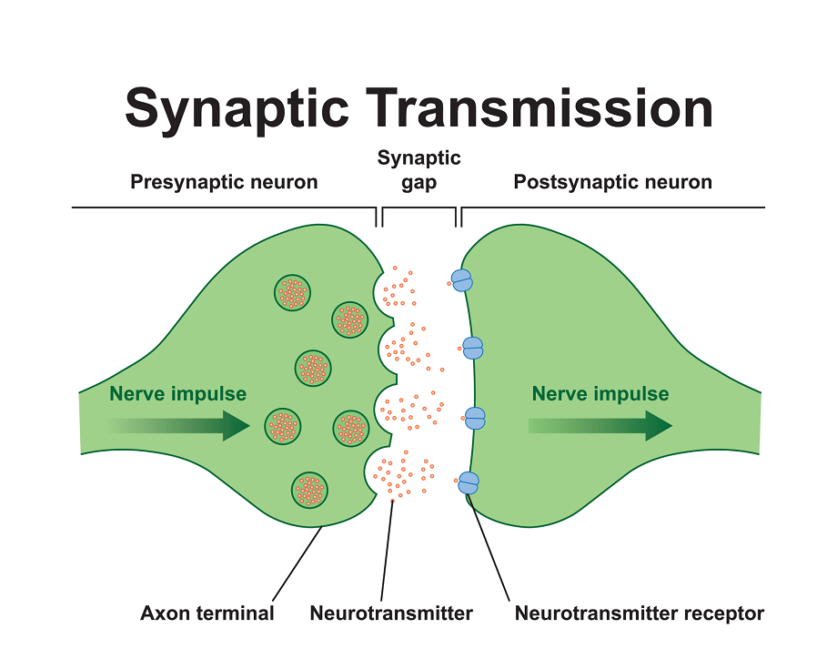
Synaptic transmission is a highly specialized process through which neurons communicate with each other or with other target cells, such as muscles or glands. This process occurs at the synapse, a junction between neurons or between a neuron and its effector target. Synaptic transmission forms the foundation of brain function, enabling processes such as perception, learning, memory, and motor coordination.
1. Definition of Synaptic Transmission
Synaptic transmission refers to the process by which one neuron transmits signals to another neuron or effector cell. This process involves the release of neurotransmitters from the presynaptic neuron, their interaction with receptors on the postsynaptic neuron, and the generation of a new electrical signal in the recipient cell.
| Type of Synapse | Description |
|---|---|
| Electrical Synapse | Direct flow of ions between neurons through gap junctions, allowing rapid communication. |
| Chemical Synapse | Use of neurotransmitters to bridge the synaptic cleft between neurons. |
2. Structure of the Synapse
The structure of a chemical synapse includes three main components:
| Component | Description | Example |
|---|---|---|
| Presynaptic Terminal | Contains synaptic vesicles filled with neurotransmitters. | Axon terminals of motor neurons. |
| Synaptic Cleft | A small gap (~20-40 nm) between the presynaptic and postsynaptic membranes. | Space between neurons in the hippocampus. |
| Postsynaptic Membrane | Contains receptor proteins that bind neurotransmitters and trigger a response. | Dendrites of pyramidal cells. |
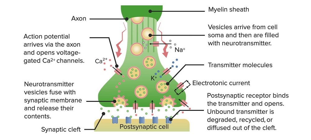
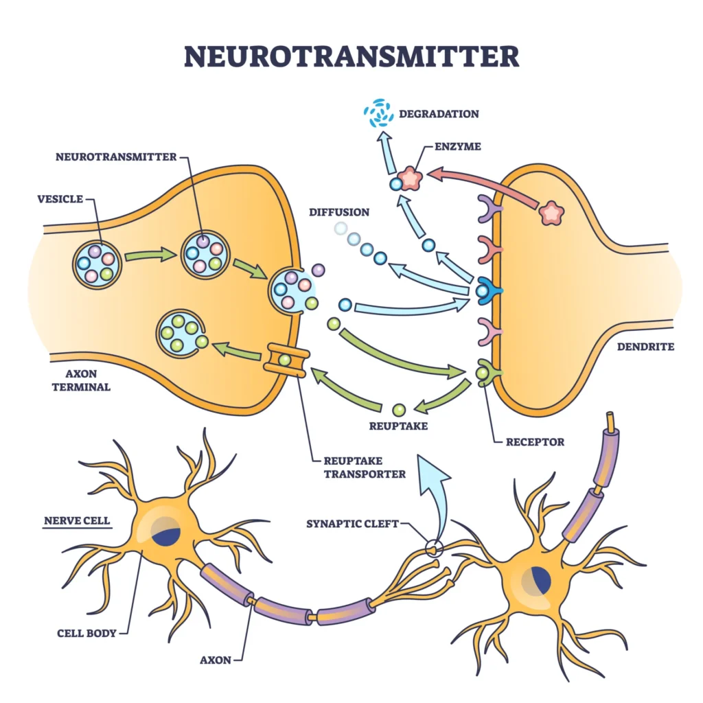
3. Phases of Chemical Synaptic Transmission
Chemical synaptic transmission involves the release of neurotransmitters from the presynaptic neuron and their binding to receptors on the postsynaptic neuron.
| Phase | Description | Key Elements |
|---|---|---|
| 1. Synthesis | Neurotransmitters are synthesized in the neuron’s soma or axon terminals. | Enzymes, precursor molecules. |
| 2. Storage | Neurotransmitters are stored in synaptic vesicles within the axon terminals. | Vesicle proteins, neurotransmitters (e.g., dopamine, serotonin). |
| 3. Release | When an action potential reaches the axon terminal, calcium ions trigger vesicle fusion with the membrane. | Voltage-gated calcium channels, SNARE proteins. |
| 4. Binding | Neurotransmitters diffuse across the synaptic cleft and bind to specific postsynaptic receptors. | Ionotropic and metabotropic receptors. |
| 5. Termination | Neurotransmitter action is terminated via reuptake, enzymatic degradation, or diffusion. | Reuptake transporters, enzymes like acetylcholinesterase. |
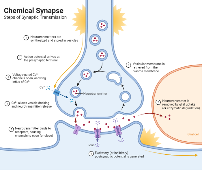
3.1. Neurotransmitter Release and Receptor Binding
Key Steps:
- Action potential triggers calcium influx in the presynaptic neuron.
- Synaptic vesicles release neurotransmitters into the synaptic cleft.
- Neurotransmitters bind to postsynaptic receptors, causing changes in the postsynaptic membrane’s potential.
| Mechanism | Description |
|---|---|
| Exocytosis | Vesicles release neurotransmitters into the synaptic cleft. |
| Ionotropic Receptors | Ligand-gated ion channels directly alter the ion flow (e.g., GABA-A receptors). |
| Metabotropic Receptors | Activate G-proteins and second messengers, leading to slower but prolonged effects (e.g., dopamine receptors). |
Research Insight:
Neurotransmitter dynamics have been implicated in psychiatric disorders. For example, abnormal serotonin transmission is linked to depression (Mössner et al., 2000).
3.2. Mechanism of Action
Figure: Illustration of neurotransmitter release, diffusion, and receptor binding (not provided here but should ideally include labeled components like vesicles, receptors, and ion channels).
| Step | Key Players | Action |
|---|---|---|
| 1 | Action potential | Depolarizes the presynaptic membrane. |
| 2 | Calcium ions | Influx through voltage-gated calcium channels triggers vesicle fusion. |
| 3 | Synaptic vesicles | Release neurotransmitters into the cleft via exocytosis. |
| 4 | Neurotransmitters | Bind to postsynaptic receptors, opening ion channels or activating G-proteins. |
| 5 | Enzymes and transporters | Break down or reabsorb neurotransmitters, clearing the synapse. |
4. Key Neurotransmitters and Functions
| Neurotransmitter | Function | Example of Action |
|---|---|---|
| Acetylcholine | Facilitates muscle contraction and memory. | Controls skeletal muscle movements; mediates learning in the hippocampus. |
| Dopamine | Regulates reward, motivation, and mood. | Influences addiction behaviors and motor control (e.g., Parkinson’s disease). |
| GABA | Main inhibitory neurotransmitter. | Prevents overexcitation of the brain, reducing anxiety. |
| Glutamate | Main excitatory neurotransmitter. | Drives synaptic plasticity and memory formation. |
| Serotonin | Modulates mood, sleep, and appetite. | Regulates emotional states and circadian rhythms. |
5. Practical Examples and Scenarios
Example 1: Reflex Arc
Scenario: A person steps on a sharp object.
- Sensory neurons transmit pain signals to the spinal cord via chemical synapses.
- Interneurons relay signals to motor neurons.
- Motor neurons activate muscles to withdraw the foot.
| Step | Neurotransmitters Involved |
|---|---|
| Sensory Signal | Glutamate released from sensory neurons. |
| Motor Response | Acetylcholine (ACh) released at neuromuscular junctions for muscle contraction. |
Example 2: Reward System Activation
Scenario: A student solves a difficult problem.
- Dopaminergic neurons in the ventral tegmental area (VTA) release dopamine into the nucleus accumbens.
- The student experiences a sense of reward and motivation.
Research Insight:
Dopamine release in the reward pathway is associated with reinforcement learning (Schultz et al., 1997).
6. Disorders and Abnormal Synaptic Function
| Disorder | Cause | Impact on Synaptic Transmission | Symptoms |
|---|---|---|---|
| Parkinson’s Disease | Dopaminergic neuron degeneration. | Reduced dopamine at synapses. | Motor impairment, tremors. |
| Alzheimer’s Disease | Accumulation of amyloid-beta plaques. | Impaired cholinergic synaptic transmission. | Memory loss, cognitive decline. |
| Epilepsy | Excessive synaptic firing. | Hyperexcitation at synapses. | Seizures. |
Research Insight:
Cholinergic therapies (e.g., acetylcholinesterase inhibitors) have shown modest benefits in Alzheimer’s patients (Birks & Harvey, 2006).
7. Role of Synaptic Plasticity
Synaptic plasticity is the ability of synapses to strengthen or weaken over time. It underlies learning, memory, and adaptation.
| Type | Description | Example |
|---|---|---|
| Long-Term Potentiation (LTP) | Persistent strengthening of synapses after repeated stimulation. | Hippocampal LTP for memory formation. |
| Long-Term Depression (LTD) | Persistent weakening of synapses after low-frequency stimulation. | Cerebellar LTD for motor learning. |
8. Disorders and Synaptic Dysfunction
| Disorder | Synaptic Dysfunction | Symptoms |
|---|---|---|
| Parkinson’s Disease | Loss of dopamine neurons in the substantia nigra. | Tremors, rigidity, bradykinesia. |
| Schizophrenia | Dysregulation of dopamine and glutamate transmission. | Hallucinations, delusions, cognitive deficits. |
| Myasthenia Gravis | Autoimmune attack on acetylcholine receptors at the neuromuscular junction. | Muscle weakness, fatigue. |
| Epilepsy | Imbalance between excitatory (glutamate) and inhibitory (GABA) transmission. | Seizures, loss of consciousness. |
9. Research Insights
- Role of Synaptic Plasticity in Memory:
- Hebb’s Rule (1949): “Cells that fire together, wire together.”
- Long-Term Potentiation (LTP): Persistent strengthening of synaptic connections underlies learning and memory formation (Bliss & Collingridge, 1993).
- Synaptic Transmission and Neurodevelopmental Disorders:
- Autism spectrum disorders involve alterations in synaptic proteins like neurexins and neuroligins (Sudhof, 2008).
- Artificial Synapses in Neural Interfaces:
- Research into bioelectronic interfaces mimics synaptic transmission to restore lost sensory or motor functions (Yang et al., 2021).
10. Conclusion
Synaptic transmission is a cornerstone of neural communication, underlying everything from basic reflexes to complex cognition. Advances in understanding synaptic mechanisms have shed light on numerous neurological and psychiatric disorders, paving the way for targeted interventions. Future research in synaptic plasticity, neurodegenerative diseases, and neural interfaces holds the promise of transformative therapeutic breakthroughs.
References
- Bliss, T. V., & Collingridge, G. L. (1993). A synaptic model of memory: Long-term potentiation in the hippocampus. Nature, 361(6407), 31-39.
- Sudhof, T. C. (2008). Neuroligins and neurexins link synaptic function to cognitive disease. Nature, 455(7215), 903-911.
- Yang, C., Guo, X., & Lu, W. (2021). Bio-inspired artificial synapses for neural interfaces. Advanced Materials, 33(21), 2007527.